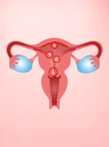
FIBROIDS,often called uterine myomas are benign smooth muscle neoplasm that typically originate from the myometrium. Its incidence is 20-30% of women over 30 years of age.
Based on location fibroids can be classified as – Submucous, Intramural, Subserous and Cervical. According to FIGO classification it has been classified as Type 0 – pedunculated intracavity
Type1 -< 50% Intramural
Type2 – > 50% Intramural
Type 3 – 100% Intramural& endometrial contact
Type 4 – 100% Intramural
Type 5 –Subserosal&>=50%Intramural
Type 6 – Subserosal&< 50% Intramural
Type 7 – Subserosal pedunculated
Whereas, ADENOMYOSIS is characterized by uterine enlargement caused by ectopic nests of endometrium located deep within the myometrium. It can be either Diffuse adenomyosis; when nests scattered throughout the myometrium or, Focal Adenomyosis ; when present as localized nodular collection.
On the basis of SYMPTOMS-
FIBROIDS can be asymptomatic or can present with bleeding, pelvic pain, pressure symptoms,or infertility( fibroids can diminish fertility but only 1-3 % cases are solely due to fibroids).On pelvic examination, uterine enlargement, irregular contour or both can be present.
Whereas ADENOMYOSIS presents with menorrhagia, dysmenorrhea, metrorrhagia, chronic pelvic pain and dyspareunia.Infertility is occasionally reported but its incidence increases with increasing age.On pelvic examination, diffusely enlarged, tender, “boggy” uterus is suggestive of adenomyosis. Severe endometriosis might present as a fixed, tender uterus, with palpable nodules within the posterior cul-de-sac and/or lining the uterosacral ligaments and rectovaginal septum.
For diagnosis of Fibroids,
USG is the imaging modality of choice. Uterine fibroids mostly appear as concentric, solidhypo- to hyper echoic depending on the ratio of smooth muscle to connective tissue and whether there is degeneration. Calcifications appear hyperechoic which either rim the tumor or scattered throughout the mass. They may have anechoic components resulting from necrosis. If they are small and isoechoic relative to the uterus, the only ultrasonographic sign may be a bulge in the uterine contour.On Doppler study, a peripheral circumferential rim of vascularity from which a few vessels arise to penetrate into the centre of the tumor is seen. MRI has an important role in assessing disease in patients in whom ultrasound findings are confusing. It allows more accurate assessment of the size, number, and location of leiomyomas, which may help identify appropriate candidates for hysterectomy alternatives such as myomectomy or UAE. Fibroids appear as areas of low to intermediate signal intensity with sharp margins on T1- and T2-weighted MRI scans.
Whereas for ADENOMYOSIS,MRI has been reported to be the most reliable diagnostic method with a reported sensitivity between 70-100% and a specificity of 86-93%. However, ultrasonography is mostly preferred because it is more widely available and significantly more cost-effective for patients.On transvaginal ultrasonography, common features include anterior or posterior myometrial wall appearing thicker than its counterpart, myometrial texture heterogeneity, small myometrial hypoechoic cysts.
Other feature includes striated projections extending from endometrium into the myometrium, ill-defined endometrial echo and a globally enlarged uterus. On Doppler studies diffuse vascularity may be seen in affected myometrium.On T2-weighted MRI, the junctional zone is more easily visualized as a low signal intensity zone adjacent to the endometrium. On T2- weighted MRI, usually a junctional zone thickness greater than 12mm is considered diagnostic for adenomyosis. Additionally, heterotopic endometrial tissue appears as small foci of increased high signal intensity within the junctional zone.
Coming to TREATMENT
In case of FIBROIDS, in asymtomatic cases, watchful waiting is the best option. Symptomatic cases can be treated medically or surgically.
For Medical management following drugs are used
• NSDS decreases dysmenorrhoea
• COCcauses decreased menstrual bleeding and 17% decrease in the risk of leiomyoma growth.
• Progestogens decreases menstrual bleeding in up to 70% cases, inducing amenorrhea in up to 30%,and may decrease uterine volume in up to 50%.
• LNG IUS reduces bleeding in 99% cases, and decreases uterine volume in about 40% .
• GnRH-analog causes uterine volume reduction in up to 50%.
• SPRM decreases bleeding in up to 98% cases, decreases fibroid volume in up to 53%.
(NSD: Non steroid anti inflammatory drugs,COC: Combined oral contraceptive, LNG IUS: Levonorgestrel releasing intrauterine system, , GnRH a: Gonadotropin releasing hormone analog, SPRM: Selective progesterone receptor modulators)
Radiologic interventions for management of fibroid include-
• Uterine artery embolization- for women having significant symptoms despite medical management and who might otherwise be considered a candidate for hysterectomy or myomectomy, and
• Magnetic Resonance –guided Focused ultrasound (MRgFUS)
Surgical management includes,
• Myomectomy, which is fertility preserving surgery, and can be performed hysteroscopically, laparoscopically,or via laparotomy.
• Hysterectomy.
For Treatment of Adenomyosis-
For improvement of symptoms, main objective of treatment is to relieve pain and bleeding.NSDS and COCS can be used for this. LNG-IUS is also effective for treatment of adenomyosis related bleeding. GnRH agonists are effective choice for women with adenomyosis related subfertility.Hysterectomy is definitive treatment.
Articles
2023


World AIDS Vaccine Day 2023: Can HIV & AIDS affect fertility or your infant’s health?
World AIDS Vaccine Day is observed every year on the 18th of May to create awa...
2023


Male Infertility Infertility Tips
Hyperspermia: Causes, Symptoms, Diagnosis & Treatment
What is Hyperspermia? Hyperspermia is a condition where an individual produ...


Guide to infertility treatments Infertility Tips
पीआईडी: पेल्विक इनफ्लैमेटरी डिजीज और निःसंतानता
पीआईडी - पेल्विक इनफ्लैमेटरी �...
2022


Infertility Tips Uterine Fibroids
Endometrial Polyps (Uterine Polyps)
What are Endometrial Polyps (Uterine Polyps)? Endometrial polyps, often ref...
2022


Female Infertility Infertility Tips
Why do You Need Fertility Treatment
As we all know infertility rate is constantly rising in our society day by day...
2022


Cesarean Section Vs Natural Birth
Surrogacy centers in Delhi and Infertility centers in Pune state that there ar...
2022


ನಿಮಗೆ ಹುಟ್ಟಲಿರುವ ಮಗುವನ್ನು ಅರ್ಥಮಾಡಿಕೊಳ್ಳುವುದು: ಗರ್ಭದಲ್ಲಿ ಮಗು ಹೇಗೆ ಬೆಳೆಯುತ್ತದೆ!
ವೀರ್ಯವು ಮೊಟ್ಟೆಯನ್ನು ಭೇಟಿಮಾಡ�...
2022


Diet Chart for Pregnant Women: The Right Food for Moms-To-Be
Pregnancy Food Chart 1. The daily diet must include the right amount of pro...
2022


Can i become pregnant while my tubes are tied?
Pregnancy is one of the most important phases in women’s life and is conside...
Pregnancy Calculator Tools for Confident and Stress-Free Pregnancy Planning
Get quick understanding of your fertility cycle and accordingly make a schedule to track it


